Heart Screening Tests and Procedures: A Complete Guide
Cardiac screening tests help detect heart conditions early often before any symptoms appear. This guide covers every major test used in heart screening, how each one works, and who should consider getting tested.
6+
Diagnostic tests covered
5 min
Average ECG duration
Non-invasive
Most screening tests
Early
Detection saves lives
What is cardiac screening?
Cardiac screening is a structured set of non-invasive diagnostic evaluations that assess the health, structure, and electrical function of the heart. Unlike diagnostic testing done after symptoms appear, screening is proactive it identifies risk factors, structural abnormalities, and rhythm disorders before they cause serious complications.
Heart disease remains one of the leading causes of premature death globally. Many conditions, including hypertension, arrhythmias, and early-stage coronary artery disease, develop silently over years. Cardiac screening provides a clinical window into cardiovascular health that lifestyle observation alone cannot offer.
Types of heart screening tests
A cardiac evaluation typically includes one or more of the following tests, chosen based on your age, symptoms, and risk profile. Each test examines a different dimension of heart health.
Electrocardiogram (ECG)
The electrocardiogram, commonly called an ECG or EKG, is one of the most widely used cardiac diagnostic tools. It records the electrical signals that travel through the heart with each heartbeat using small electrode patches placed on the skin of the chest, arms, and legs.
What it detects
- Arrhythmias and irregular heart rhythms
- Previous or ongoing heart attacks (myocardial infarction)
- Heart enlargement (hypertrophy)
- Conduction abnormalities
- Ischemia (reduced blood supply to heart muscle)
Procedure details
- Duration: 5–10 minutes
- Non-invasive, painless
- No special preparation required
- Results available immediately
- Performed at rest (resting ECG)
2D Echocardiogram
A 2D echocardiogram uses high-frequency sound waves (ultrasound) to create real-time images of the heart. It is the primary imaging tool for evaluating cardiac structure and function, allowing clinicians to directly observe how the heart is built and how well it pumps.
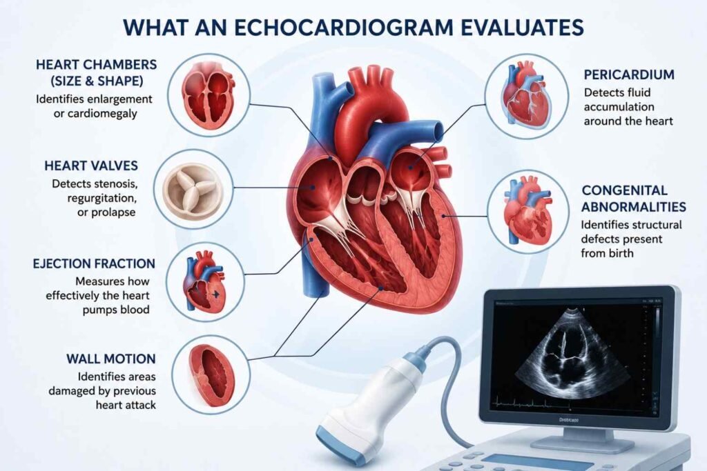
The procedure involves applying a water-based gel to the chest and moving a transducer probe across the skin to capture images. It is entirely painless and typically takes 30–45 minutes. A 2D echo is especially important for patients with heart murmurs, unexplained breathlessness, or a family history of structural heart disease.
Treadmill Test (TMT) -Exercise Stress Test
The treadmill test, also called an exercise stress test, monitors how the heart performs under physical exertion. Since some cardiac conditions only manifest during activity, the TMT provides diagnostic information that a resting ECG cannot capture.
How it works
You walk or run on a treadmill at progressively increasing speeds and incline levels. Heart rate, blood pressure, and ECG readings are continuously monitored throughout the test. The test is stopped when the target heart rate is achieved or if symptoms develop.
Who it’s recommended for
Individuals with chest pain or breathlessness during physical activity, those with known coronary artery disease, patients being evaluated before cardiac rehabilitation, and individuals with multiple cardiovascular risk factors.
A positive TMT result where the heart shows signs of stress-induced ischemia usually prompts further investigation such as a coronary angiogram. The test is contraindicated in patients with acute heart conditions or severe resting hypertension.
Holter Monitoring
Holter monitoring involves wearing a small portable ECG device continuously for 24 to 48 hours (sometimes up to 7 days). Unlike a standard ECG which records only a few seconds of heart activity, a Holter monitor captures the heart’s electrical activity throughout an entire day including sleep, meals, and physical activity.
Holter monitoring is particularly valuable for detecting intermittent arrhythmias that may not appear during a brief clinical ECG — such as paroxysmal atrial fibrillation, ventricular ectopics, or unexplained palpitations.
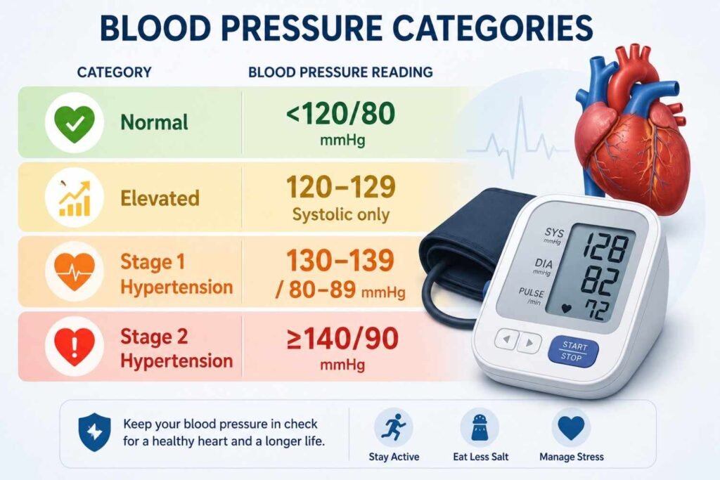
Blood Pressure Assessment
Blood pressure assessment is the most fundamental component of any cardiac screening protocol. Hypertension (high blood pressure) is both a major risk factor for heart disease and a direct cause of arterial damage, left ventricular hypertrophy, and stroke.
Regular blood pressure monitoring is recommended for all adults regardless of age. Ambulatory blood pressure monitoring (ABPM) where readings are taken automatically over 24 hours provides a more accurate clinical picture than a single office reading.
Who needs heart screening?
Screening is most valuable for individuals with elevated cardiovascular risk, but it also benefits those who want a proactive baseline assessment of heart health.
- Adults over 40 years of age cardiovascular risk increases significantly with age
- Individuals with known high blood pressure or diabetes
- People with high cholesterol or a history of lipid disorders
- Current or former smokers
- Those with a family history of heart disease, heart attack, or sudden cardiac death
- Individuals with obesity or metabolic syndrome
- Anyone experiencing chest pain, palpitations, shortness of breath, or unexplained fatigue
How to prepare for heart screening tests
General preparation
- Wear comfortable, loose-fitting clothing
- Bring all current medication details
- Bring previous cardiac reports if available
- Avoid heavy meals immediately before tests
- Inform staff of any recent symptoms
For treadmill test (TMT)
- Fast for 3–4 hours before the test
- Avoid caffeine on the day of the test
- Wear comfortable sports shoes
- Inform cardiologist of any chest pain history
- Certain medications may be paused consult your doctor
Conclusion
Understanding heart screening tests and procedures is the first step toward taking control of your cardiovascular health. From a quick resting ECG that captures your heart’s electrical rhythm to a 2D echocardiogram that visualises its structure, a treadmill test that reveals how your heart responds under stress, and a Holter monitor that records every beat across an entire day each test serves a distinct and valuable diagnostic purpose. Together, these evaluations give clinicians a comprehensive picture of your heart’s health, helping detect conditions early when they are most treatable. Whether you have existing risk factors such as high blood pressure, diabetes, or a family history of heart disease, or simply want a proactive baseline assessment, cardiac screening remains one of the most reliable tools available for protecting long-term heart health. If you are considering heart screening, consult a qualified cardiologist to determine which tests are appropriate for your age, symptoms, and risk profile, and take that important step toward a healthier heart today.
For evidence-based guidelines on heart disease prevention and cardiovascular wellness, visit the American Heart Association – a globally trusted resource for patients and healthcare professionals alike.
FREQUENTLY ASKED QUESTIONS
1. What are the most common heart screening tests?
The most common heart screening tests include the Electrocardiogram (ECG), 2D Echocardiogram, Treadmill Test (TMT), Holter Monitoring, Blood Pressure Assessment, and Doppler Ultrasound Studies. Each test evaluates a different aspect of cardiovascular health from electrical rhythm and structural integrity to blood flow and stress response. A cardiologist will recommend the appropriate combination based on your age, symptoms, and risk factors.
2. Is heart screening the same as a cardiac checkup?
Not exactly. A cardiac checkup is a broader consultation that includes a physical examination, medical history review, and discussion of symptoms and lifestyle. Heart screening refers specifically to the diagnostic tests performed during or as part of that checkup. In practice, most comprehensive cardiac checkups include one or more screening tests such as an ECG or echocardiogram to provide objective clinical data alongside the consultation.
3. Can heart screening detect a future heart attack?
Heart screening cannot predict a heart attack with certainty, but it can identify risk factors and early warning signs that significantly increase the likelihood of one occurring. Tests like the Treadmill Test can reveal stress-induced ischemia, while blood pressure and cholesterol assessments identify arterial risk factors. Early detection through regular screening allows doctors to intervene with medication, lifestyle changes, or further investigation before a cardiac event occurs.
4. How long does a full heart screening take?
The duration depends on which tests are included. A resting ECG takes approximately 5 to 10 minutes, a 2D echocardiogram takes around 30 to 45 minutes, and a Treadmill Test typically takes 30 to 60 minutes including preparation and recovery time. If multiple tests are performed in a single visit, a comprehensive heart screening session may take between one and three hours in total. Holter monitoring extends over 24 to 48 hours as it is worn continuously throughout daily activity.
5. At what age should I start getting heart screening tests?
Most cardiologists recommend a baseline cardiac evaluation starting around the age of 30 to 40, particularly if any risk factors are present such as high blood pressure, diabetes, obesity, or a family history of heart disease. For individuals with no known risk factors, routine heart health checks during annual physical examinations are generally considered sufficient until the age of 40, after which more structured cardiac screening becomes increasingly important.
6. Are heart screening tests covered under health insurance?
Coverage varies depending on your insurance provider and policy terms. Many health insurance plans in India cover diagnostic tests like ECG and echocardiogram when they are prescribed by a registered physician. Preventive health checkup packages, which often include cardiac screening, are also covered under several policies. It is advisable to check with your insurance provider or the diagnostic facility beforehand to understand what is included in your plan.
7. What is the difference between a 2D echo and a colour Doppler echo?
A standard 2D echocardiogram produces two-dimensional ultrasound images of the heart’s structure its chambers, walls, and valves showing size and movement. A colour Doppler echocardiogram adds colour-coded blood flow information on top of those images, allowing cardiologists to see the direction and velocity of blood moving through the heart in real time. Colour Doppler is particularly useful for diagnosing valve disorders such as regurgitation or stenosis where blood flow patterns are abnormal.
8. Can I eat before a heart screening test?
This depends on the specific test being performed. For a resting ECG, blood pressure assessment, or echocardiogram, eating beforehand is generally not a concern. However, for a Treadmill Test (TMT), it is recommended to fast for at least three to four hours and avoid caffeine on the day of the procedure, as a full stomach or stimulants can affect heart rate and test accuracy. Always follow the specific instructions provided by your healthcare provider when booking your screening appointment.
9. Can young and healthy individuals benefit from heart screening?
Yes. While heart screening is most urgently recommended for individuals with known risk factors, young and apparently healthy individuals can also benefit from a baseline evaluation. Some congenital heart conditions, inherited arrhythmias, and structural abnormalities present no obvious symptoms but can be identified through screening. Athletes and individuals engaging in high-intensity physical activity are also often advised to undergo cardiac screening to rule out conditions such as hypertrophic cardiomyopathy before starting intense training programmes.
10. What happens if an abnormality is found during heart screening?
If a screening test reveals an abnormality, your cardiologist will discuss the findings with you and recommend appropriate next steps. Depending on the nature of the finding, this may include additional diagnostic tests such as a coronary CT angiogram or stress echocardiogram, a referral to a specialist, or the initiation of medication and lifestyle modifications. An abnormal result does not always indicate a serious condition many findings are minor and manageable with early intervention. The purpose of screening is precisely to catch such issues at a stage where they can be effectively addressed.